
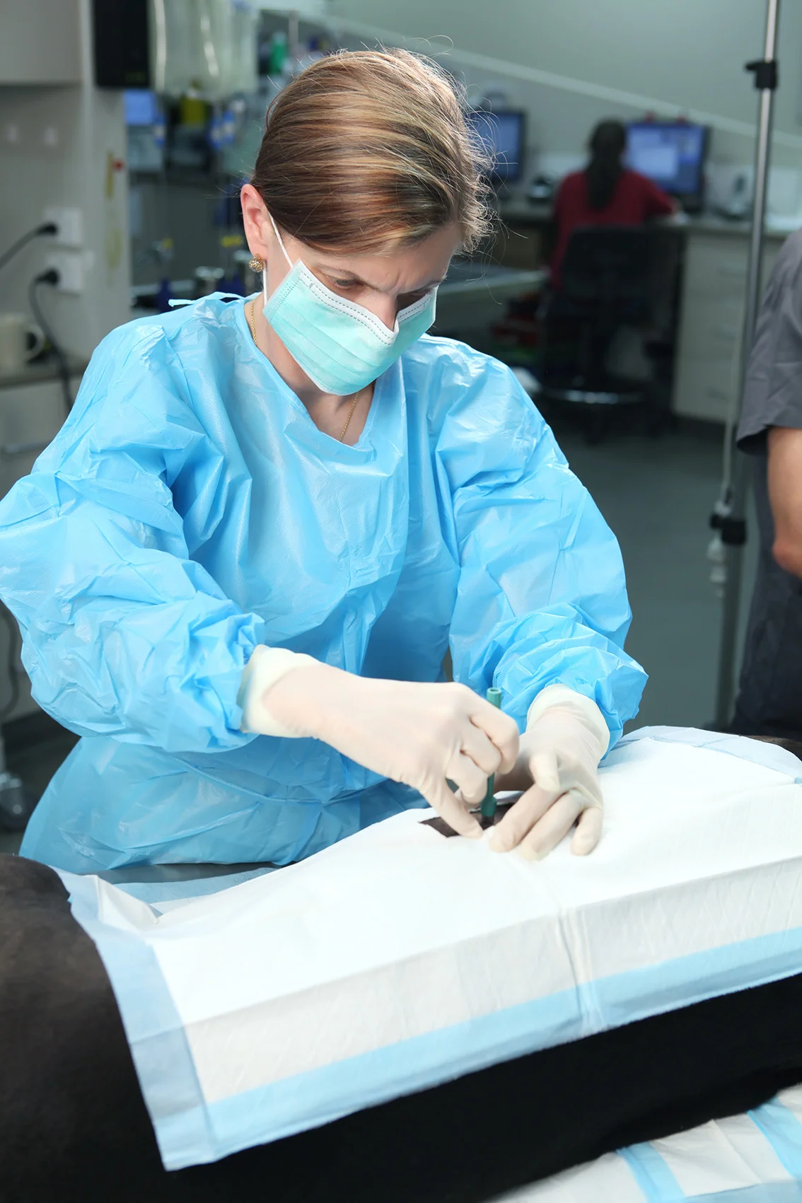
Skin Biopsies & Histopathology
Some skin conditions can be diagnosed through examination, skin samples and other tests. Others require us to take a closer look at the structure of the skin itself.
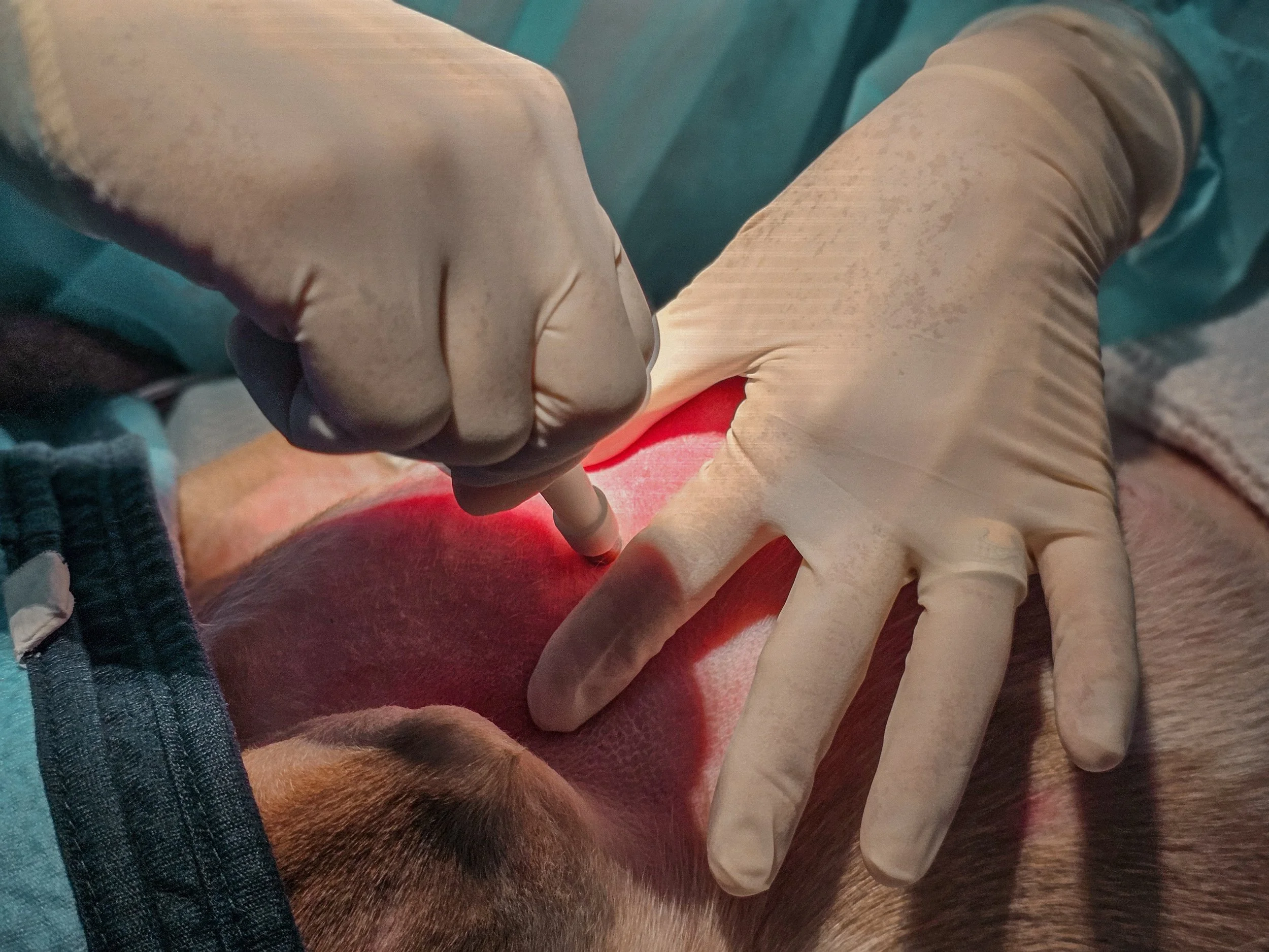
A skin biopsy involves removing one or more small samples of affected skin. These samples are then examined under a microscope by a veterinary pathologist, a process called histopathology.
Histopathology can provide valuable information about what is happening within the different layers and structures of the skin and, for some conditions, is an important step towards reaching a definitive diagnosis.
Why might my pet need a skin biopsy?
Your veterinary dermatologist may recommend a biopsy when a skin condition cannot be confidently diagnosed through examination and routine testing alone.
Common reasons include:
Unusual or unexplained skin lesions
Persistent ulcers or wounds that aren't healing as expected
Lumps, growths or lesions where neoplasia (cancer) is suspected
Skin disease that isn't responding as expected to treatment
Blisters, vesicles, pustules or significant crusting
Hair and follicle disorders
Suspected immune-mediated or autoimmune skin disease
Conditions affecting the sebaceous glands
Certain nutritional or metabolic skin disorders
Any skin condition where examining the tissue architecture may help establish a diagnosis
Biopsy can be particularly useful when different skin diseases look similar on the surface but require very different treatments.
What happens during a skin biopsy?
Before taking a biopsy, your veterinary dermatologist will carefully examine your pet's skin and select the areas most likely to provide useful diagnostic information.
Usually, several small samples are taken rather than just one. Skin disease can vary from one area to another, so selecting the right lesions and obtaining appropriate samples is an important part of achieving a useful result.
Depending on the location and number of samples required, the procedure may be performed using local anaesthesia with sedation or under general anaesthesia. Your dermatologist will determine the most appropriate approach for your pet.
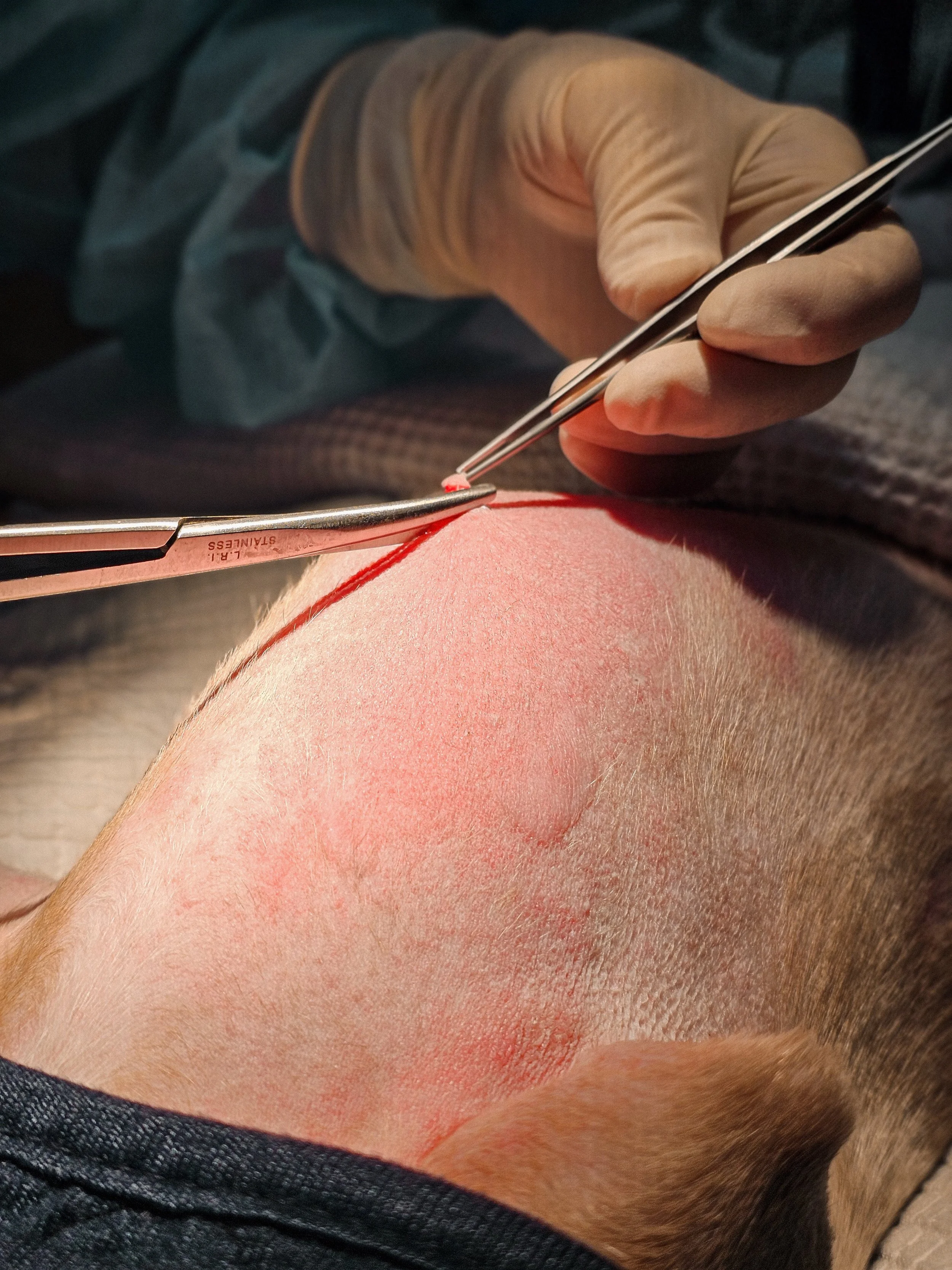
A small biopsy instrument is used to remove a sample containing the layers of skin required for examination. Most biopsy sites are then closed with one or two sutures.
The samples are carefully prepared and submitted for histopathological examination.
Why does a veterinary dermatologist perform the biopsy?
With skin biopsies, where and when the sample is taken can be just as important as taking the sample itself.
Different dermatological diseases produce characteristic changes in particular types and stages of lesions. Older, scratched, infected or previously treated areas may sometimes provide less useful information than carefully selected fresh lesions.
Veterinary dermatologists are trained to recognise these patterns and select appropriate biopsy sites based on the conditions being considered.
We also provide the pathologist with your pet's relevant clinical history and our dermatological findings. Histopathology is most valuable when the microscopic changes can be interpreted alongside the complete clinical picture.
What happens to the biopsy samples?
The samples are preserved and sent to a veterinary pathology laboratory, where very thin sections of the tissue are prepared, stained and examined under a microscope.
The veterinary pathologist assesses the cells, hair follicles, glands and other structures within the skin, looking for patterns of inflammation, infection, abnormal cell growth and other changes that may help identify the disease process.
Once the histopathology report is available, your veterinary dermatologist interprets the findings together with your pet's history, examination and other test results.
Sometimes histopathology provides a definitive diagnosis. In other cases, it helps us narrow down the possibilities or determine what further investigation may be required.
What should I expect after a skin biopsy?
Most biopsy sites are very small and heal quickly.
Your pet may have several small sutured areas where samples were collected. We'll provide instructions for caring for these sites at home, which may include keeping them clean and dry and preventing your pet from licking, scratching or rubbing them.
Complications following skin biopsy are uncommon, although minor bleeding, swelling, infection or delayed wound healing can occasionally occur. Certain medications and underlying health conditions can also affect healing.
What happens when the results come back?
Getting the pathology report is only part of the process.
Your veterinary dermatologist will interpret the biopsy findings in the context of your pet's complete dermatological assessment and explain what the results mean, whether further investigation is required, and what treatment or management options are recommended.
Our aim is not simply to name the skin condition, it's to use that diagnosis to develop the most appropriate plan for your pet.
Worried about your pet? Give us a call. We treat your pet like one of our own. Call now on: +61 08 6277 9430 (Murdoch) and (Balcatta) or email: dermnurses@adcperth.com